
Digital X-Ray Department
Digital X-Ray imaging supports the evaluation of bones, joints and other structures and plays an important role in orthopaedic diagnosis.
Clear
Imaging
Faster
Diagnosis
Better
Planning
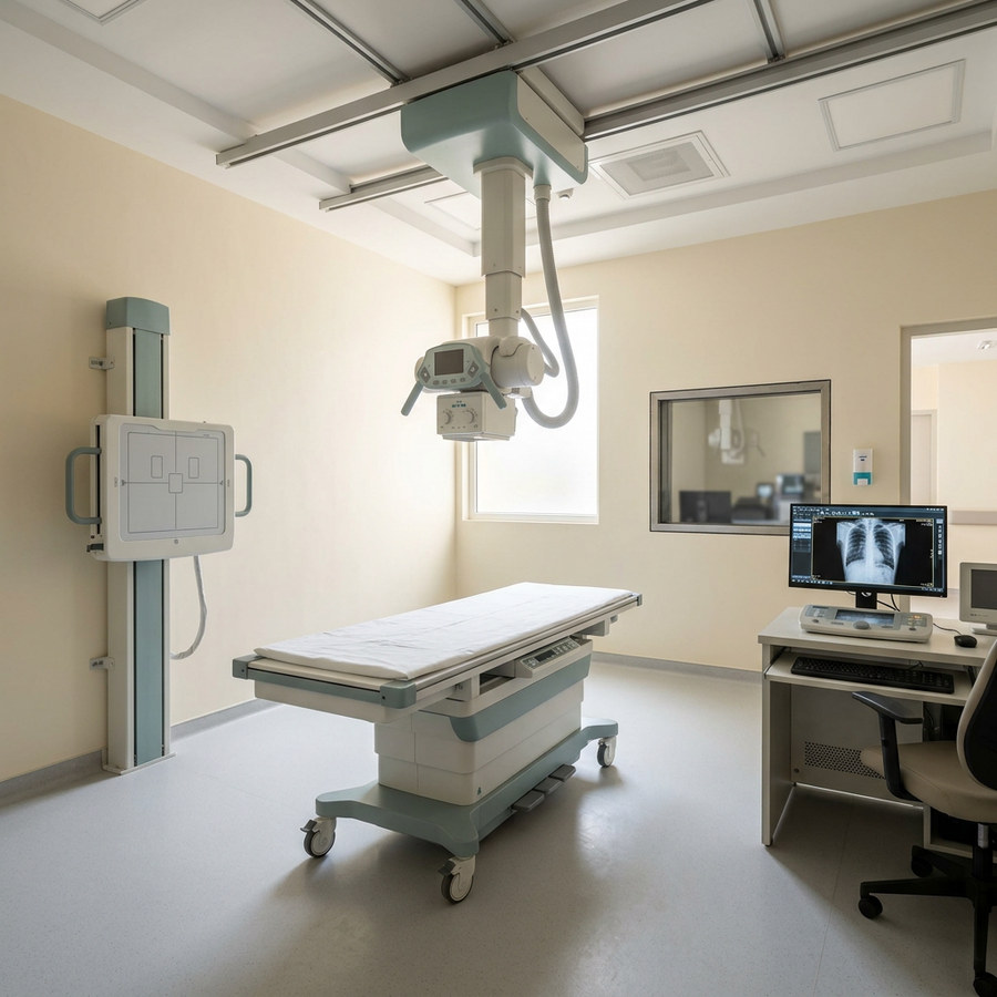
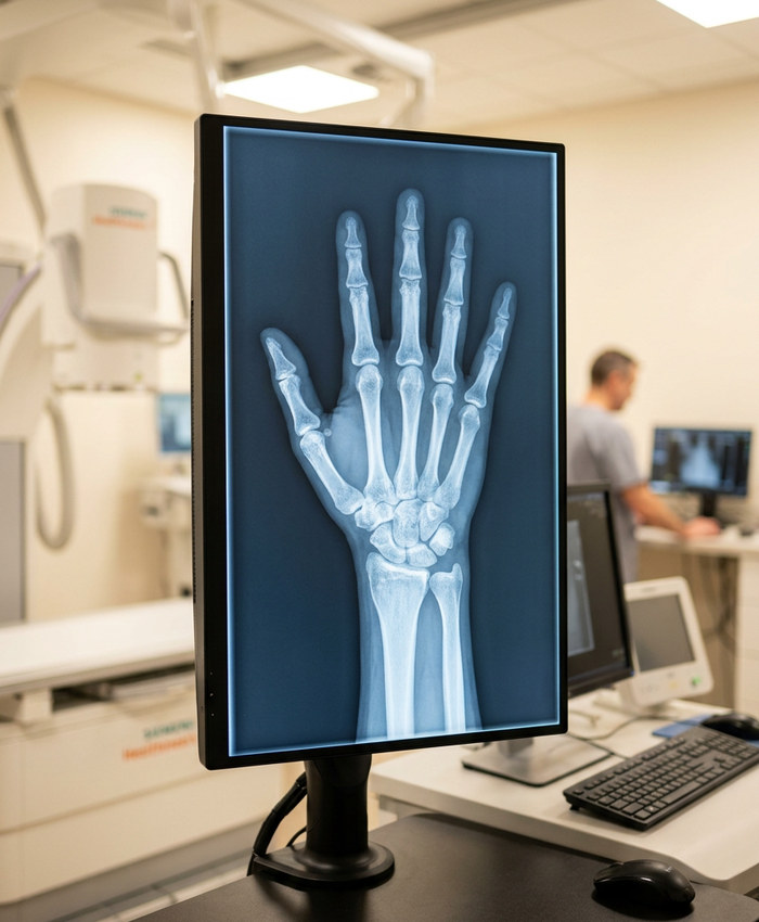
What is Digital X-Ray?
Digital X-Ray uses X-ray technology to create electronic images that can be reviewed by healthcare professionals for diagnostic assessment.
Digital images support orthopaedic diagnosis and follow-up, helping the clinical team plan appropriate treatment.
“Clear images. Confident decisions.”
Imaging for Accurate Diagnosis
Digital X-ray support for orthopaedic evaluation and follow-up
Clear Images, Faster Answers
Digital X-Ray imaging supports the evaluation of bones, joints and other structures, and plays an important role in orthopaedic diagnosis.
Images are electronically processed and available for review by the clinical team, supporting timely treatment planning.
Common Applications
Fracture Detection
Imaging to identify fractures and their pattern.
Joint Evaluation
Assessment of joints and their alignment.
Bone Alignment
Evaluation of bone position and structure.
Follow-Up Imaging
Tracking healing progress over time.
Pre-Operative Assessment
Imaging support before planned procedures.
How Digital X-Ray Works
A four-step process from positioning to clinical review.
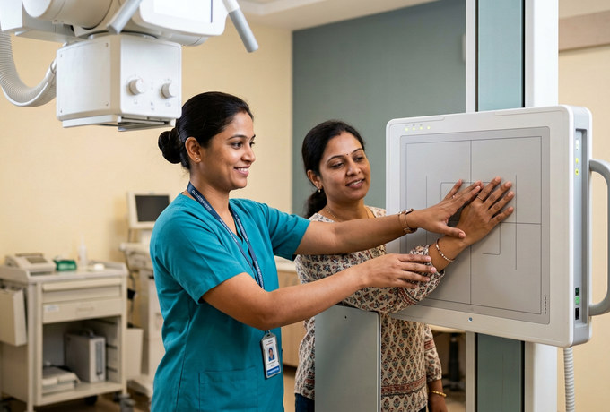
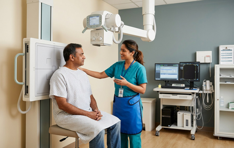
01. Positioning
The patient is positioned appropriately.
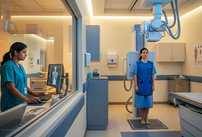
02. Image Capture
The required X-ray images are obtained.
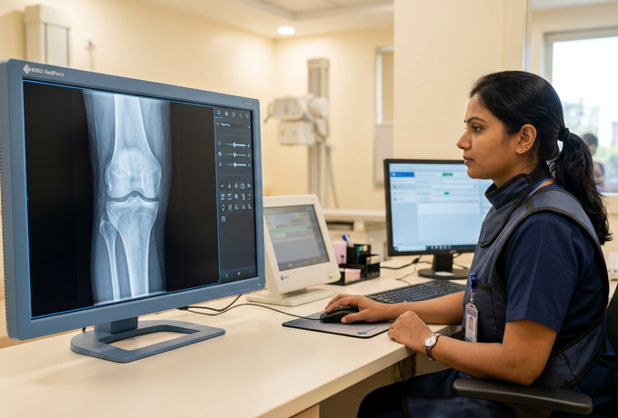
03. Digital Processing
Images are electronically processed.

04. Clinical Review
The healthcare team reviews the images for treatment planning.
Benefits of Digital X-Ray
Clear imaging that supports confident diagnosis.
Clear Digital Images
Electronically processed images support accurate review.
Efficient Availability
Images are available promptly for the clinical team.
Fracture Assessment
Imaging support for evaluating fractures and healing.
Follow-Up Evaluation
Useful for tracking progress over time.
Patient Safety & Care
X-Ray examinations are performed using appropriate radiation-safety practices and only when clinically indicated.
The radiology team follows established safety practices in handling images, and exposure is kept to the level required for a clear diagnostic result.
Why Choose Shri Datta?
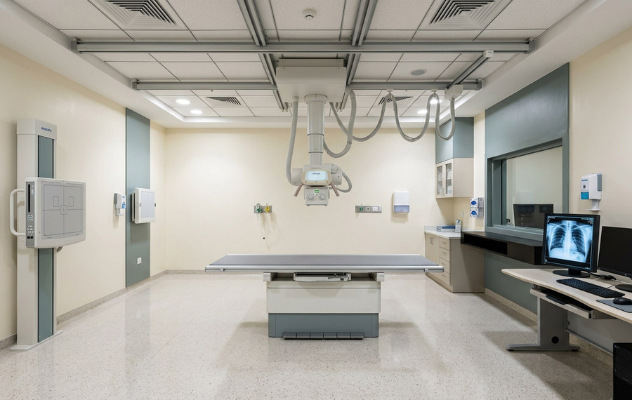
Our imaging team supports the orthopaedic department with clear, digitally processed X-ray images for diagnosis and follow-up.
Efficient image availability helps the clinical team plan and review treatment with confidence, reducing waiting time for patients.
Frequently Asked Questions
Answers to key questions regarding our digital X-ray services.

Clear Imaging. Faster Diagnosis.
Digitally processed X-ray images that support confident orthopaedic diagnosis and treatment planning.
- Digital Imaging
- Radiology Team
- Follow-Up Support